
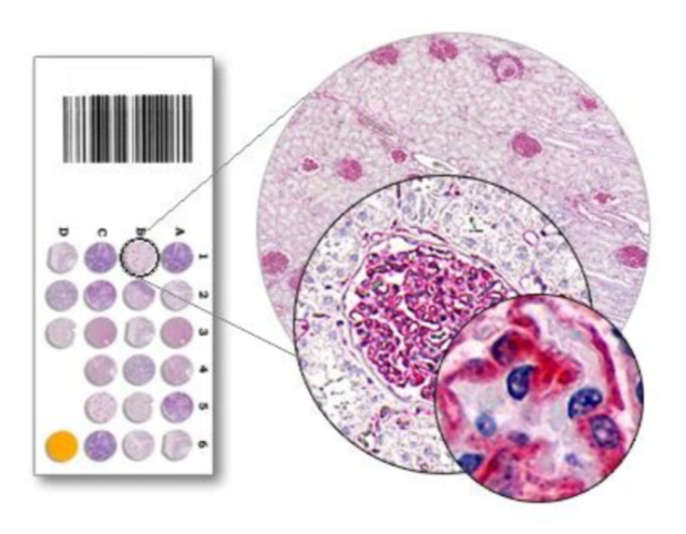
A tissue section is a thin slice of biological tissue prepared for microscopic examination. Tissue sectioning is a fundamental step in histology, pathology, medical research, and diagnostic laboratories. By preparing tissue sections correctly, scientists and medical professionals can study cell structures, detect diseases, and understand biological processes at the microscopic level tissue array.
This article provides a comprehensive overview of tissue sections, including their definition, preparation methods, types, common techniques, tools used, applications, and challenges. Whether you are a student, researcher, or healthcare professional, understanding tissue sectioning is essential for accurate microscopic analysis.
What Is a Tissue Section?
A tissue section is a very thin slice of tissue mounted on a glass slide for examination under a microscope. These sections are typically only a few micrometers thick, allowing light or electrons to pass through them for detailed visualization.
Tissue sections are commonly used in:
- Histology
- Pathology
- Biomedical research
- Medical diagnostics
- Veterinary science
- Forensic analysis
By analyzing a tissue section, professionals can observe cellular organization, identify abnormalities, and make diagnostic decisions.
Importance of Tissue Sectioning in Histology
Tissue sectioning plays a critical role in histology because tissues are naturally too thick to examine directly under a microscope. Proper tissue sectioning ensures:
- Clear visualization of cells and structures
- Accurate diagnosis of diseases
- Consistent and reproducible results
- High-quality staining and imaging
Without well-prepared tissue sections, even the most advanced microscopes cannot produce meaningful results.
Steps in Tissue Section Preparation
Preparing a tissue section involves several carefully controlled steps. Each step affects the final quality of the section and the accuracy of the analysis.
1. Fixation
Fixation preserves the tissue structure and prevents decay. Common fixatives include:
- Formalin
- Paraformaldehyde
- Glutaraldehyde
Fixation stabilizes proteins and cellular components, maintaining tissue integrity.
2. Dehydration
After fixation, water is removed from the tissue using graded alcohol solutions. This step is necessary for embedding the tissue in a solid medium.
3. Clearing
Clearing replaces alcohol with a solvent such as xylene, making the tissue transparent and compatible with embedding materials.
4. Embedding
Embedding provides structural support to the tissue. Common embedding media include:
- Paraffin wax
- Resin (for ultra-thin sections)
- OCT compound (for frozen sections)
5. Sectioning
The embedded tissue is sliced into thin sections using specialized instruments such as microtomes or cryostats.
6. Mounting and Staining
The tissue section is placed on a glass slide and stained to enhance contrast. Common stains include hematoxylin and eosin (H&E).
Types of Tissue Sections
There are several types of tissue sections, each suited for specific applications and analysis techniques.
Paraffin Tissue Sections
Paraffin-embedded tissue sections are the most commonly used in routine histology and pathology.
Advantages:
- Long-term storage
- Good structural preservation
- Compatible with many staining techniques
Disadvantages:
- Longer preparation time
- Some antigen loss during processing
Frozen Tissue Sections
Frozen tissue sections are prepared using a cryostat without extensive processing.
Advantages:
- Rapid preparation
- Better preservation of enzymes and lipids
- Useful for intraoperative diagnosis
Disadvantages:
- Lower structural quality
- Shorter storage life
Resin Tissue Sections
Resin-embedded tissue sections are used for high-resolution studies.
Advantages:
- Extremely thin sections
- Excellent cellular detail
- Suitable for electron microscopy
Disadvantages:
- Complex preparation
- More expensive materials
Semi-Thin and Ultra-Thin Sections
- Semi-thin sections (0.5–1 µm) are used for light microscopy.
- Ultra-thin sections (50–100 nm) are essential for transmission electron microscopy.
Tools Used for Tissue Sectioning
High-quality tissue sections depend on precision instruments designed for controlled slicing.
Microtome
A microtome is used to cut thin tissue sections from paraffin or resin blocks. Types include:
- Rotary microtome
- Sliding microtome
- Ultramicrotome
Cryostat
A cryostat is a refrigerated microtome used for frozen tissue sections. It maintains low temperatures to preserve tissue integrity during sectioning.
Blades and Knives
- Steel blades
- Disposable blades
- Glass knives
- Diamond knives (for ultra-thin sections)
Each blade type is selected based on tissue hardness and desired thickness.
Common Staining Techniques for Tissue Sections
Staining enhances contrast and highlights specific tissue components.
Hematoxylin and Eosin (H&E)
The most widely used stain for tissue sections:
- Hematoxylin stains nuclei blue
- Eosin stains cytoplasm pink
Special Stains
Special stains target specific structures, such as:
- Periodic Acid–Schiff (PAS) for carbohydrates
- Masson’s trichrome for connective tissue
- Silver stains for nerve fibers
Immunohistochemistry (IHC)
IHC uses antibodies to detect specific proteins in a tissue section. This technique is essential in cancer diagnosis and research.
Applications of Tissue Sections
Tissue sections are used across many scientific and medical fields.
Medical Diagnosis
Pathologists examine tissue sections to diagnose:
- Cancer
- Inflammatory diseases
- Infections
- Autoimmune disorders
Biopsies rely heavily on accurate tissue sectioning for correct diagnosis.
Biomedical Research
Researchers use tissue sections to:
- Study disease mechanisms
- Analyze gene expression
- Evaluate drug effects
- Understand tissue development
Education and Training
Medical and biology students learn anatomy, histology, and pathology by studying tissue sections under microscopes.
Veterinary and Forensic Science
Tissue sections are used in:
- Animal disease diagnosis
- Toxicology studies
- Cause-of-death investigations
Common Challenges in Tissue Sectioning
Despite its importance, tissue sectioning presents several challenges.
Section Artifacts
Artifacts may occur due to:
- Improper fixation
- Dull blades
- Incorrect cutting speed
Common artifacts include folds, tears, and compression.
Tissue Brittleness or Softness
- Over-dehydrated tissue becomes brittle
- Under-processed tissue may be too soft to cut
Balancing processing conditions is crucial for high-quality tissue sections.
Thickness Variability
Inconsistent section thickness can affect staining and interpretation. Proper instrument calibration is essential.
Best Practices for High-Quality Tissue Sections
To ensure reliable results:
- Use sharp, clean blades
- Maintain proper fixation times
- Control temperature and humidity
- Handle tissue gently during processing
- Regularly maintain sectioning equipment
Following standardized protocols improves reproducibility and diagnostic accuracy.
Future Trends in Tissue Sectioning
Advancements in technology continue to improve tissue section preparation.
- Automated tissue processors
- Digital pathology and whole-slide imaging
- Cryo-electron microscopy
- AI-assisted tissue analysis
These innovations enhance precision, efficiency, and diagnostic confidence.
Conclusion
A tissue section is a cornerstone of histology, pathology, and biomedical research. From fixation and embedding to sectioning and staining, each step plays a vital role in producing high-quality slides for microscopic analysis. Understanding the types, techniques, tools, and applications of tissue sections allows professionals to achieve accurate diagnoses, meaningful research outcomes, and reliable educational results.
As technology advances, tissue sectioning continues to evolve, offering greater precision and deeper insights into the microscopic world. Mastery of tissue section preparation remains essential for anyone working in life sciences and medical fields.